MICROSCOPE
.jpeg)
.jpeg)
.jpeg)
https://www.google.com/search?sxsrf=ALeKk02h79EMJgW5c_vFNhop6-3uZCqgfw%3A1624568561655&source=univ&tbm=isch&q=About%20MICROSCOPE&fir=24rc-mtgX6oyuM%252C33KOEOu2rW8WuM%252C_%253Bp23q3J5mf3WtoM%252C7qsL_FRjkwwjOM%252C_%253BHByh1ey9kjHCRM%252CihgP1dEwSU0csM%252C_%253BaUgNUaYwbhytNM%252CvbH_v6AQeB2bAM%252C_%253Bw4NAlW3b6woExM%252Cr7dKrYdJ-HKbIM%252C_%253Br7QyyWo6fSKLZM%252CTZ8aQV48NvA-EM%252C_%253BgsxxVy5vTBOQ5M%252CzEffoz8zYNdNAM%252C_%253B9m7zHkQ5t873hM%252CdxO0odM29lzn2M%252C_%253B8GLWDMM-zjoqaM%252CA-KBe8vhxkqOMM%252C_%253BulpAuury1s7H3M%252CcUqKbWLEow477M%252C_%253B0AGYky6fWlM9XM%252C2_FfYhylyDZRAM%252C_&usg=AI4_-kQaIvJovK8z7-shKMT6nDe_SoTvDA&ved=2ahUKEwjDt-nElbHxAhXKm2oFHTpaANgQv7IFegQIARAG&biw=393&bih=675#imgrc=HByh1ey9kjHCRM
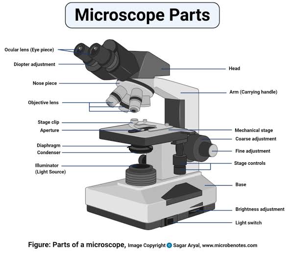
Stricture of a light microscope
To study small organisms and cells we use a microscope. The most common microscope is the light or compound microscope.
A light microscope has two set of glass lenses: the objective lenses and the eyepiece lenses. Most microscope have two or three objective attached to a rotating nose piece. These lenses usually are 5x,10x, 20x, 40x, and 100x. (A 10x lens magnifies the image of the objective lens. Its magnification ranges from 5x to 15x.
The magnifying power of microscope is the product of the magnifications of the objective and the eyepiece: if the eyepiece magnifies 15x and the objective 40x, the total magnification is 15x 40=600x.
Resolution of a microscope
A microscope not only magnifies the image but also separates minute details. the ability of a microscope to distinguish two very close objects as being separated from each other is its resolution or resolving power. Thus, if we look at two objects which are close together through a low resolution microscope, we see them as a single object. However, through a high resolution microscope we can distinguish the two objects. The resolution power is about half the wavelength of light (approximately 250 NM).Therefore, the resolving power of a light microscope is limited. At the most, it can distinguish two point which are 250nm (0.25 um) apart and magnife about 1 500 times. Since most cell organelles are smaller than 200nm, they cannot be seen through a light microscope.
Note microscope objects are measured in micros. A micros is 1/1000mm. The symbol for micron is the Greek letter u (mu) which is 1/1000000mm.
How to use the microscope
Step 1: Clip the specimen slide on the stage of the microscope. Make sure the specimen is over the hole in the stage.
Step 2: Rotate the nose piece to bring the low power objective lens just above the specimen.
Step 3: place a lamp in front of the microscope and tilt the microscope mirror so that the light is directed up through the eyepiece.
Step 4: look through the eyepiece and adjust the diaphragm of the iris until the light is bright, but not dazzling.
Step 5: look from the side of the microscope, and turn the coarse adjustment knob and lower the objective lens until it is close to the slide.
Step 6: look through the eyepiece and slowly turn the coarse adjustment knob and raise the one Ken to focus the specimen sharply.
To look at the specimen under high power, Rotate the nose until the high power objective lens is over the specimen. Then, look through the eyepiece and carefully focus the specimen, using the fine adjustments knob. Adjust the illumination if necessary
Making a wet mount of a specimen
Specimen for viewing through a microscope are usually prepared as thin slices or secrions. Soft tissue like root tips are squashed, while liquid tissue like blood is smeared. Transparent part of a specimen are offer stained with appropriate dyes to make them visibe. The prepared specimen is then mounted on a glass microscope slide as follows:
Step 1: place a drop of the mounting liquid (usually water or glycerine) in the central of a clean dry microscope slide and put the specimen in this liquid
Step 2: stand a cover slip on the side at an angle of 45°. Pull it along the slide until it touches the drop of liquid. Using a mounting needle, very carefully, lower the cover slip until it is flat on the top of the liquid drop. With a piece of filter paper, draw off, if any , excess mounting liquid around the cover slip.
Electron microscope
Many organelles in cells are too small to be viewed through a light microscope. An electron microscope is used to study such objects. It used a beam of electron whose wavelength is very much shorter than that of light. the beam is focused by powerful electromagnets.the image produced cannot be viewed directly. Onto a fluorescent screen, from which black and white photographs of the image called photoelectron micrographs are taken.
An electron microscope has a resolving power of around 1nm and magnifies objects over 500 000 times. Thus, it can give a far more detailed picture of the structures in a cell than the most powerful light microscope.
First atomic force microscope
^ Characterization and Analysis of Polymers. Hoboken, NJ: Wiley-Interscience. 2008. ISBN 978-0-470-23300-9.^ Bardell, David (May 2004). "The Invention of the Microscope". BIOS. 75 (2): 78–84. doi:10.1893/0005-3155(2004)75<78:tiotm>2.0.co;2. JSTOR 4608700.^ The history of the telescope by Henry C. King, Harold Spencer Jones Publisher Courier Dover Publications, 2003, pp. 25–27 ISBN 0-486-43265-3, 978-0-486-43265-6^ Atti Della Fondazione Giorgio Ronchi E Contributi Dell'Istituto Nazionale Di Ottica, Volume 30, La Fondazione-1975, p. 554^ a b Murphy, Douglas B.; Davidson, Michael W. (2011). Fundamentals of light microscopy and electronic imaging (2nd ed.). Oxford: Wiley-Blackwell. ISBN 978-0-471-69214-0.^ Sir Norman Lockyer (1876). Nature Volume 14.^ Albert Van Helden; Sven Dupré; Rob van Gent (2010). The Origins of the Telescope. Amsterdam University Press. pp. 32–36, 43. ISBN 978-90-6984-615-6.^ "Who Invented the Microscope?". Retrieved 31 March 2017.^ Eric Jorink (2010-10-25). Reading the Book of Nature in the Dutch Golden Age, 1575-1715. ISBN 978-90-04-18671-2.^ William Rosenthal, Spectacles and Other Vision Aids: A History and Guide to Collecting, Norman Publishing, 1996, pp. 391–92^ Raymond J. Seeger, Men of Physics: Galileo Galilei, His Life and His Works, Elsevier – 2016, p. 24^ J. William Rosenthal, Spectacles and Other Vision Aids: A History and Guide to Collecting, Norman Publishing, 1996, page 391^ uoregon.edu, Galileo Galilei (Excerpt from the Encyclopedia Britannica)^ Gould, Stephen Jay (2000). "Chapter 2: The Sharp-Eyed Lynx, Outfoxed by Nature". The Lying Stones of Marrakech: Penultimate Reflections in Natural History. New York: Harmony. ISBN 978-0-224-05044-9.^ a b Wootton, David (2006). Bad medicine: doctors doing harm since Hippocrates. Oxford [Oxfordshire]: Oxford University Press. p. 110. ISBN 978-0-19-280355-9.[page needed]^ Liz Logan (27 April 2016). "Early Microscopes Revealed a New World of Tiny Living Things". Smithsonian.com. Retrieved 3 June 2016.^ Knoll, Max (1935). "Aufladepotentiel und Sekundäremission elektronenbestrahlter Körper". Zeitschrift für Technische Physik. 16: 467–475.^ Goldsmith, Cynthia S.; Miller, Sara E. (2009-10-01). "Modern Uses of Electron Microscopy for Detection of Viruses". Clinical Microbiology Reviews. 22 (4): 552–563. doi:10.1128/cmr.00027-09. ISSN 0893-8512. PMC 2772359. PMID 19822888.^ Morita, Seizo (2007). Roadmap of Scanning Probe Microscopy. Berlin, Heidelberg: Springer-Verlag Berlin Heidelberg. ISBN 978-3-540-34315-8.^ a b c d e f g h i j Lodish, Harvey; Berk, Arnold; Zipursky, S. Lawrence; Matsudaira, Paul; Baltimore, David; Darnell, James (2000). "Microscopy and Cell Architecture". Molecular Cell Biology. 4th Edition.^ a b c d e f g Alberts, Bruce; Johnson, Alexander; Lewis, Julian; Raff, Martin; Roberts, Keith; Walter, Peter (2002). "Looking at the Structure of Cells in the Microscope". Molecular Biology of the Cell. 4th Edition.^ "The Nobel Prize in Chemistry 2014 – Scientific Background" (PDF). www.nobelprize.org. Retrieved 2018-03-20.^ "The Nobel Prize in Chemistry 2014". www.nobelprize.org. Retrieved 2018-03-20.^ a b Erko, A. (2008). Modern developments in X-ray and neutron optics. Berlin: Springer. ISBN 978-3-540-74561-7.^ Pennycook, S.J.; Varela, M.; Hetherington, C.J.D.; Kirkland, A.I. (2011). "Materials Advances through Aberration-Corrected Electron Microscopy" (PDF). MRS Bulletin. 31: 36–43. doi:10.1557/mrs2006.4.^ Aspden, Reuben S.; Gemmell, Nathan R.; Morris, Peter A.; Tasca, Daniel S.; Mertens, Lena; Tanner, Michael G.; Kirkwood, Robert A.; Ruggeri, Alessandro; Tosi, Alberto; Boyd, Robert W.; Buller, Gerald S.; Hadfield, Robert H.; Padgett, Miles J. (2015). "Photon-sparse microscopy: visible light imaging using infrared illumination" (PDF). Optica. 2 (12): 1049. Bibcode:2015Optic...2.1049A. doi:10.1364/OPTICA.2.001049. ISSN 2334-2536.^ a b Bhushan, Bharat, ed. (2010). Springer handbook of nanotechnology (3rd rev. & extended ed.). Berlin: Springer. p. 620. ISBN 978-3-642-02525-9.^ Sakurai, T.; Watanabe, Y., eds. (2000). Advances in scanning probe microscopy. Berlin: Springer. ISBN 978-3-642-56949-4.^ "Quantum Microscope for Living Biology". Science Daily. 4 February 2013. Retrieved 5 Febr